


the health strategy
management and technology review
for in-person health, digital health,
and hybrid health strategy
Joaquim Cardoso MSc.
Senior Research and Strategy Officer (CRSO),
Chief Editor and Senior Advisor
January 18, 2023
Key takeaways
Complex aortic repairs are lengthy operations requiring frequent in-procedure imaging, which exposes patients and staff to radiation.
A new imaging device called Fiber Optic RealShape (FORS) at the University of Texas (UT) Southwestern is making complex aortic repairs safer for patients and operating room staff.
- FORS uses light to visualize blood vessels, significantly reducing the need for X-rays during minimally invasive vascular procedures.
- The safety of patients and staff is a priority, making the reduction of radiation exposure during procedures crucial.
UT Southwestern was one of the select medical centers participating in the early implementation of FORS.
- FORS employs hair-thin optical fibers in specially designed catheters and wires to display its position and shape within the body.
- Strain on the optical fibers changes the light’s pathway, enabling a computer algorithm to reconstruct and visualize the full shape of the device in real-time, providing surgeons with a 3D view of the blood vessel.
- Surgeons can overlay the FORS-generated view on computed tomography images, creating a roadmap for surgery, and reducing the need for X-rays.
The use of FORS is expected to expand to other vascular procedures in the future, potentially becoming a standard technology for various cardiovascular procedures.
DEEP DIVE
Safer imaging technology for complex aortic repairs uses light instead of X-rays
Vascular News
22nd August 2022
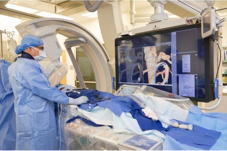
A new imaging device at University of Texas (UT) Southwestern (Dallas, USA) is making complex aortic repairs safer for patients and operating room staff by dramatically reducing their exposure to radiation. The device, known as Fiber Optic RealShape (FORS; Philips), uses light to visualise blood vessels, nearly eliminating the need for X-rays typically used during minimally invasive vascular procedures.
“
Complex aortic repairs tend to be long operations that require frequent in-procedure imaging. Every time a surgeon presses on the X-ray pedal, the patient and staff including assistants, nurses, scrub techs, anaesthetists, and X-ray techs gets a dose of radiation,” said Carlos Timaran, professor of surgery at UT Southwestern. “The safety of each of these individuals is paramount, so reducing radiation exposure during these procedures is an important goal.”
UT Southwestern was one of about a dozen medical centres in the USA and Europe chosen to participate in the early implementation of FORS. Timaran has completed more than 300 fenestrated endovascular aortic repairs as part of his physician-sponsored investigational device exemption study, in which a patient-specific graft is used to support the aorta and its main branches.
As an alternative to conventional imaging, the FORS device uses light travelling through hair-thin optical fibres built in specially designed catheters and wires to display its position and shape inside the body. Once this device is placed within a blood vessel, the strain on the optical fibres changes the light’s pathway. By analysing how light reflects back along the fibres, a computer algorithm reconstructs and visualises the full shape of the device. The result is a real-time, three-dimensional view of the blood vessel that surgeons can overlay on computed tomography images taken before the procedure, providing a roadmap that surgeons can view in any angle to guide the surgery. Timaran said that far fewer X-rays are necessary when using FORS, significantly reducing patients’ and staff’s exposure.
He expects FORS use to expand to other vascular procedures over time. “This technology could potentially be used for any cardiovascular procedure,” he said. “This will eventually be the goal.”
Originally published at https://vascularnews.com on August 22, 2022.
Names mentioned
Carlos Timaran, professor of surgery at UT Southwestern